How to read biopsy results and understand the results
Biopsy results
During a biopsy, 10 to 12 samples of prostate tissue are removed. These are then sent to pathology for histopathological examination under a microscope. The histological examination then establishes the actual diagnosis of prostate cancer. Processing of the samples usually takes 7-10 days but may take longer in some cases.
The pathologist, doctor processing the prostate biopsy samples, describes the characteristics of each sample, whether it is a benign growth of the prostate glands (benign hyperplasia), an inflammatory process or a prostate tumour.
Establishing the correct classification is essential for the subsequent choice of the appropriate treatment modality. The disease severity increases with a higher number for each TNM letter.
Based on the extent of the disease as determined by the TNM classification, the histological grade of the maturing tumour (Gleason score, GS) and the PSA concentration before treatment, prostate tumours are divided into three basic groups – low, intermediate and high risk, which also determine the likely course of the disease (prognosis) and help in selecting the most appropriate treatment for the patient.
| PROGNOSIS | |
| Risk group | Characteristics |
| Low risk | T1-T2a and GS 2-6 and PSA < 10 ng/ml |
| Intermediate risk | T2b-T2c or GS 7 or PSA 10-20 ng/ml |
| High risk | T3a or GS 8-10 or PSA > 20 ng/ml |
| Very high risk | risk T3b-T4 N0 or any T N1 or any T any N M1 |
TNM classification and disease stage
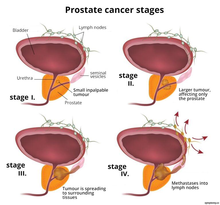
Once a diagnosis of prostate cancer has been made, the extent of the disease (stage of disease) must also be determined. This is based on the results of a rectal examination of the prostate and some special investigations – PSA level, pelvic CT, pelvic MRI, skeletal scintigraphy, lung X-ray, PET/CT scan (not all of which are necessary).
- In a localised disease, the tumour affects only the prostate and no spread outside the prostate is detected (no metastases). There is a distinction between early-stage prostate cancer – the tumour is localised inside the prostate and the prostate capsule is not affected, and locally advanced prostate cancer – in which the prostate capsule is affected by the tumour and it can spread to the immediate surroundings of the prostate.
- In generalised disease, metastatic spread of the tumour is detected. The most common targets of metastasis are the pelvic lymph nodes and the skeleton. However, it can (although not nearly as often) spread to other organs.
The TNM classification is used to simply describe the extent of the tumour and determine the stage of the disease. The stage of the disease is one of the criteria based on which the doctor determines the treatment. Accurate diagnosis of the disease stage is crucial for further treatment. In particular, it is critical to determine whether the tumour has spread beyond the prostate. That is, whether metastases have also formed (most often in the bones or vertebrae of the spine). Describing the extent of the disease is called staging and the letters indicate the following:
- T: primary tumour – in the case of prostate cancer T0 means that no tumour is present, T1 to T4 denotes a specific description of the extent of the tumour, its size and/or its relationship to surrounding structures
- N: regional lymph nodes – N0 = lymph nodes not affected by tumour, N1 to N3 = description of the affect on the lymph nodes and the extent of such affect
- M: metastases – M0 means that distant metastases are not present, M1 distant metastases are present
Numbers are further assigned to each letter, in the case of prostate cancer, T 1-4 is used for the letter T and indicates its clinical stage:
- Stage T1 – this is a clinically undetectable tumour, neither by palpation nor by imaging. This stage is further subdivided and indicated by lower case letters.
Stage T1a indicates that the tumour has been detected incidentally by histology and is present in ≤ 5% of the tissue.
Stage T1b indicates that the tumour was incidentally detected histologically and is present in > 5% of the tissue.
Stage T1c indicates that the tumour has been detected on punch biopsy (indicated e.g. by an elevated PSA level).
- Stage T2 – this is the stage where the tumour only affects the prostate.
Stage T2a indicates that the tumour affects half of one lobe of the prostate or less.
Stage T2b indicates that the tumour affects more than one half of one prostate lobe but not both.
Stage T2c indicates that the tumour affects both lobes.
- Stage T3 – the tumour has spread beyond the prostate capsule.
Stage T3a indicates that part of the bladder is affected by the tumour.
Stage T3b indicates that the tumour also affects the seminal vesicles.
- Stage T4 – tumour affects structures other than the bladder and seminal vesicles*
*Source: https://www.uroweb.cz/index.php?pg=dg–nadory-prostaty–diagnostika
